
Our CT Service For Your Pet
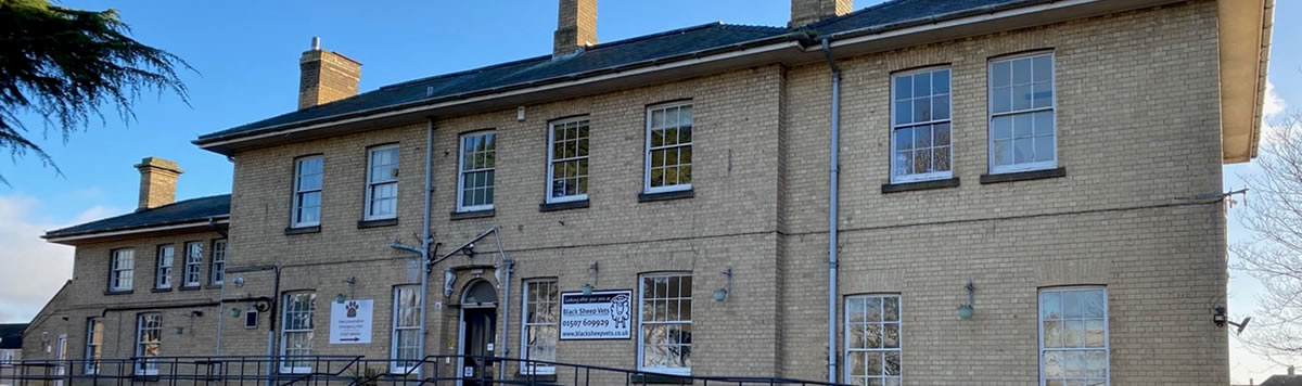
Black Sheep Vets now offers CT scanning at our practice in Louth. The service is designed for local practices who want advanced diagnostic imaging for their patients without referring them on to a distant referral hospital.
We carry out the scan, including contrast studies where indicated, and studies are reported by VetCT. The report is sent directly to your practice and the patient returns to you for ongoing management.
About CT
Computed tomography produces thin cross-sectional images of the body which can be reviewed in any plane and reconstructed in 3D. Because it removes the superimposition seen on conventional radiographs, CT is particularly valuable for complex anatomy such as the nasal cavity, tympanic bullae, skull, spine, thorax and joints.
Pre- and post-contrast studies allow assessment of vascular structures, lesion enhancement and tissue perfusion, and are used for angiography, CT urography, oncological staging and surgical planning.
All scans are performed under general anaesthesia.
Why refer to us?
Same-day scanning. With 24 hour care on site, we will always see CT referrals on the same day.
Local access. A short journey means less stress for the patient and less disruption for the owner, which is particularly helpful for fractious, geriatric or trauma patients.
Specialist reporting. All studies are reported by VetCT’s specialist radiologists. The report goes straight back to your practice, so you remain the point of contact for your client throughout.
Client continuity. Clients who come to us for a CT scan cannot register with Black Sheep Vets for other veterinary care for 4 months after their scan. This is a scan-and-return service and the case stays with you, unless you want the patient to have further treatment with us at East Lincs Emergency Vets.
Anaesthesia and recovery. Patients are monitored throughout anaesthesia and during recovery, and can stay with us overnight under 24 hour care if required.
Cases CT can help with
Nasal and sinus disease
– Chronic nasal discharge, epistaxis and sneezing
– Differentiating neoplasia, fungal rhinitis and chronic inflammatory rhinitis
– Suspected nasal foreign bodies
Ear, skull and oral cavity
– Otitis media and interna, including assessment of the tympanic bullae before surgery
– Nasopharyngeal and middle ear polyps in cats
– Skull and mandibular fractures, and temporomandibular joint disease
– Oral masses, including assessment of bone involvement and surgical planning
– Tooth root and periapical disease not adequately assessed on dental radiographs
Thorax
– Screening for pulmonary metastases, with significantly greater sensitivity than thoracic radiography for small nodules
– Unexplained coughing, dyspnoea or lung pattern not resolved on radiographs
– Lung lobe torsion, spontaneous pneumothorax and bullous lung disease
– Mediastinal, chest wall and pulmonary masses
Abdomen
– Congenital portosystemic shunts, using CT angiography to identify shunt morphology and plan surgery
– Ectopic ureters and other urinary tract abnormalities, using CT urography
– Abdominal masses, including organ of origin, vascular invasion and resectability
– Adrenal masses, including assessment for caudal vena cava invasion
– Staging of abdominal neoplasia, including regional lymph nodes
Spine
– Suspected intervertebral disc extrusion
– Vertebral fractures and luxations
– Congenital vertebral malformations
– Discospondylitis and vertebral neoplasia
Musculoskeletal
– Elbow dysplasia, including fragmented medial coronoid process and joint incongruity
– Complex, articular and pelvic fractures, including 3D reconstruction for surgical planning
– Lameness where radiographs have not identified a cause
– Bone tumours, including assessment of local extent
Oncology
– Staging before surgery, chemotherapy or radiotherapy
– Assessment of tumour margins and local invasion for surgical planning
– Monitoring response to treatment
